MRI Prostate Cancer Results Part3
Summary of Page
Impressions:
Impressions:
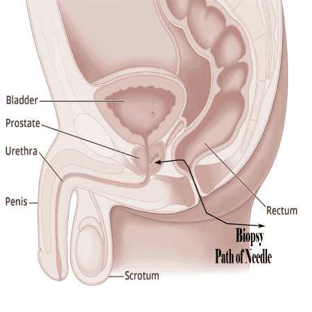
1. Lesion #1: PIRADS $. 10mm lesion left anterior peripheral zone mid base, increased in the interval.
2. Wedge-shaped: and linear areas of T2 hypointensity in the peripheralzone likely representing sequelae of prostatiis.
3. BPH: Of central gland.
4. Diverticulosis: coli.
1. Lesion #1:
Lesion #1: PI-RADS . 10mm lesion left anterior peripheral zone mid base, increased in the interval.
Excerpt:
Prostate Imaging Reporting and Data System (PI-RADS) is a structured reporting scheme for multiparametric prostate MRI in the evaluation of suspected prostate cancer in treatment naive prostate glands. This article reflects version 2.1 (v2.1), published in 2019 and developed by an internationally representative group involving the American College of Radiology (ACR), European Society of Urogenital Radiology (ESUR), and AdMeTech Foundation
2. Wedge-shaped:
Wedge-shaped: and linear areas of T2 hypointensity in the peripheralzone likely representing sequelae of prostatiis.
Excerpt:
Synopsis
This study investigated the multi-parametric MRI features and pathologic outcome of wedge shaped lesions on T2-weighted images in 76 patients. A greater percentage of wedge shaped features were found to be malignant than shown previously. Malignant wedge shaped regions were primarily highly hypointense on ADC maps and showed early enhancement on DCE-MRI. Benign wedge shaped lesions were predominantly mildly hypointense on ADC maps and showed no early enhancement and pathologically outcome showed prostatitis, hemosiderin-laden macrophages, prominent blood vessels, intraluminal blood and atrophy. Malignant wedge shaped lesions were found to have significantly lower ADC compared to benign wedge shaped regions
3. BPH:
BPH of central gland.
Excerpt:
The Benign Prostatic Hyperplasia (BPH) Symptom Score, also known as the International Prostate Symptom Score (IPSS), is a clinical tool used to assess the severity of symptoms in men with Benign Prostatic Hyperplasia (BPH). BPH is a common condition in aging men characterized by the non-cancerous enlargement of the prostate gland, which can lead to urinary symptoms such as frequent urination, weak stream, and incomplete bladder emptying. The BPH Symptom Score helps healthcare providers evaluate the severity of these symptoms, guide treatment decisions, and monitor the effectiveness of interventions over time.
Norby
4. Diverticulosis:
Diverticulosis: coli.
Diverticulosis is a common condition that can develop in your colon, especially as you get older. It means that little pouches form in the inside lining of your colon. They usually don’t cause any problems. But rarely, they may bleed or develop an infection (diverticulitis).
Helpful Information
1. Radiologist, Be Aware:
Radiologist, Be Aware: Ten Pitfalls That Confound the Interpretation of Multiparametric Prostate MRI.
Excerpt:
OBJECTIVE. In this article, we describe 10 diagnostic challenges that may confound the interpretation of multiparametric prostate MRI for tumor, grouped into three categories on the basis of our experience: normal anatomic structures that may be misinterpreted as suspicious lesions if their normal appearance is not recognized, benign processes that may mimic tumor, and technical issues relating to acquisition and interpretation of diffusion-weighted imaging that may decrease sensitivity for tumor. Strategies for addressing these challenges are suggested.
2. Can Cancer Be Detected by an MRI?
Can Cancer Be Detected by an MRI?
Excerpt:
An MRI is an effective test for detecting cancer in many parts of the body. It’s especially helpful at creating detailed images of soft tissue, including tumors. However, there are some types of cancer that it cannot detect.
Got low risk lesion?
When is a lesion a low risk?
Common clinical criteria for a low‑risk prostate lesion (typical thresholds used to offer active surveillance):
- Grade: Gleason score 6 (Grade Group 1).
- PSA: < 10 ng/mL.
- Clinical stage: cT1c–cT2a (tumor confined to prostate, limited extent).
- Tumor burden on biopsy: limited number/percentage of positive cores (exact cutoffs vary by protocol).
- PSA density (PSAD): often < 0.15 ng/mL/cm³ is used to identify very low‑risk cases.
- Concordant imaging/biopsy: mpMRI without high‑risk features; confirmatory biopsy or targeted sampling recommended before surveillance.
Hypointensity & Hyperintensity
Hypointensity & Hyperintensity
Excerpt:
to areas on MRI that appear darker than surrounding tissues, often indicating the presence of prostate cancer, while **hyperintensity** appears brighter and can indicate areas of high cellularity or restricted diffusion, which may also suggest cancer. Both terms describe how lesions are visualized on MRI scans, helping in the diagnosis and assessment of prostate cancer.
Another Biopsy needed 1.
| Another Biopsy? 05-19-2026 | ||
|---|---|---|
| What we know from MRI doc. | ||
| # | Description | Date |
| 1. | PSA 5.86 | 02-16-2026 |
| 2. | PSA Density : 0.12 ng/ml/cc. | 03-10-2026 |
| 3. | Est. volume: 47.4cc was 37.2cc | 03-10-2026 |
| 4. | Gleason score 3+4 = 7 | 02-12-2024 |
| 5. | Lesion #1: PIRADS 4. | 03-10-2026 |
| 6. | Size: 10mm was 5mm | 03-10-2026 |
| 7. | T2: Score 4. | 03-10-2026 |
| 8. | DWI/ADC: Score 3. | 03-10-2026 |
| 9. | Mean ADC value 0.655x10⁻³mm2/s | 03-10-2026 |
| 10. | DCE: Positive | 03-10-2026 |
| 11. | PIRADS score: 4 - High | 03-10-2026 |
| 12. | BPH of central gland | 03-10-2026 |
| 13. | Diverticulosis coli. | 03-10-2026 |
| Changes after the MRI | ||
| 1. | PSA 3.96 Quest Diagnostic | 05-05-2026 |
| 2. | PSA Density from 0.24-0.12-0.083 | 05-05-2026 |
Another Biopsy needed 2.
PSA doubling time (PSADT):
Shortening PSADT (e.g., months vs years) suggests more rapid tumor growth;
PSA velocity: absolute rise per year; higher velocity implies faster progression but is less used than PSADT.
Commonly used thresholds:
1. <3–6 months = very fast,
2. 6–12 months = fast,
3. 12–24 months = intermediate,
4. >24 months = slow
Note:
Now I collect all the info from the MRI, Blood tests and the latest PSA numbers. With all that info I can make a counter argument not to get another Biopsy at this time. If the PSA suddenly changes, of course I will reevaluate my decision and get another MRI and or a Biopsy. This is down the road.
Norby
Pro Con Biopsy.
Pro Biopsy:
1. make sure Cancer accgreciveness has not changed.
2. Gleason score change.
Con Biopsy:
1. PSA is getting smaller from 9.13 to 3.97
2. PSA Dencity change from 0.24 to 0.08 with PSA 3.97.
3. Another Anesthesia
4. low grade spreading the Cancer
Prostate Be Shrunk Naturally?
Benign Prostatic Hyperplasia: Can the Prostate Be Shrunk Naturally?
Excerpt:
Benign prostatic hyperplasia (BPH) is a common age-related condition that can cause urinary symptoms due to prostate growth. While natural approaches may not directly shrink the prostate, dietary changes, lifestyle measures, and plant-based therapies may help manage symptoms and support prostate health alongside conventional treatment.
Excerpts from my Diary:
Reaction from a Nurse.
The question by the nurse surprised me in a big way. The question was, What are you taking for medication without a prescription from the doctor? I was thinking what kind a question is that, my answer was Ivermectin, clear and with no hesitation. Later I realized why she was asking me that question in the first place. She noticed that my PSA number was going down from 9.3 to 5.53, that is a 40.5% reduction of the cancer. With that kind of reduction, I had to take something against the cancer. On my documentation from the hospital, I took no medication at all. So I had to take something without a prescription from my doctor. Than she asked me if it was OK to write it into my records that I was taking Ivermectin. That was OK with me at that time.
Norby
Excerpts from my Diary:
Now the doctors reaction.
Later the doctor came into the room, we made small talk, after that I told him that I took Ivermectin. His reaction was interesting. First of all he did not know that the latest PSA number came down from 9.3 to 5.53. I felt like he was indifferent to the result, meaning he did not praise the result nor did he condemn it. I do not believe him, that he did not know the PSA number, because he looked at the same documents on the computer like the nurse did, so he also saw the PSA number came down. I also gave him a copy of my writings, so he gets a better understanding of my thinking why I do not want the operation or the radiation as my first option of action.
Norby